

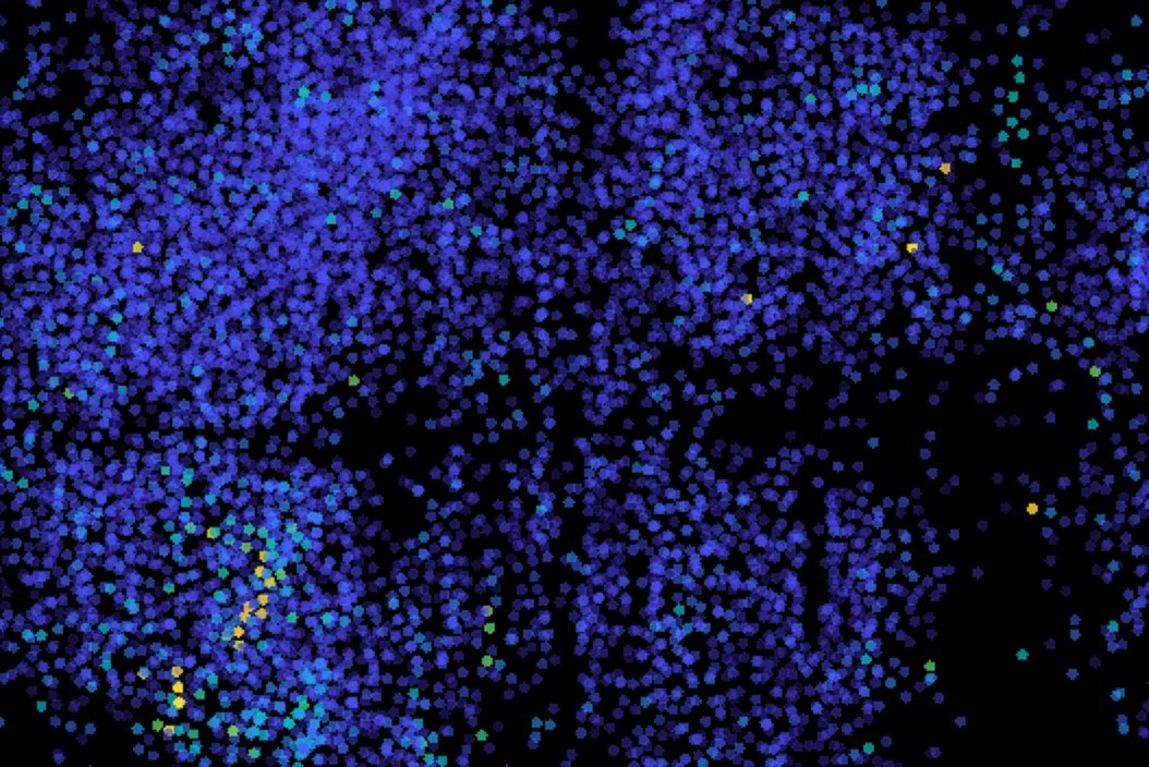
BCIwiki (bciwiki.com) — MIT engineers have adapted a light sheet microscope to image electrical activity in neurons distributed across the brain of an entire zebrafish (Danio rerio), scanning the whole brain 200 times per second, or once every five milliseconds. The work, from Ed Boyden's Synthetic Neurobiology Group at MIT, was announced on August 14, 2026 and published in Nature Methods.
Calcium imaging, often used to measure neuron activity, tracks calcium flowing into neurons after they fire and therefore serves only as a proxy; it resolves activity on the order of seconds or minutes, too slow to capture single spikes. Genetically encoded voltage indicators are fluorescent proteins expressed in neurons that fluoresce when a neuron fires, giving direct observation of voltage, but previous work could image only small, localized populations.
To reach millisecond-scale activity across a large volume, the team increased the image acquisition speed of the microscope's camera and boosted scanning speed using remote refocusing. With this approach the researchers recorded patterns of neural activity from neurons throughout the brain in response to ultraviolet light.