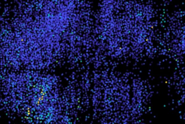
MIT engineers have adapted a light-sheet microscope to image the electrical activity of neurons across a zebrafish's entire brain, scanning the whole brain 200 times a second — once every five milliseconds. Calcium imaging, the usual proxy for neural activity, resolves activity only on the order of seconds and cannot capture single spikes, while genetically encoded voltage indicators report membrane potential directly but had previously been limited to small, localized populations. Faster camera acquisition and remote refocusing pushed volumetric imaging to rates that resolve individual neuronal impulses, revealing brain-wide activity patterns evoked by ultraviolet light; the study appears in Nature Methods.