
/ EN

The University of Melbourne

3 entries
September 2026

Synchron Stent Electrode Beats Scalp EEG per Channel in One ALS Patient

A participant with severe upper-limb paralysis caused by amyotrophic lateral sclerosis (ALS) wore an endovascular stent-electrode array and a scalp EEG cap at the same time, in the same session, while attempting ankle flexion and extension. Both recordings changed markedly during attempted movement, but the stent array showed stronger per-channel motor modulation and was unaffected by skull attenuation. Scalp EEG was more susceptible to eye blinks and jaw-muscle activity, while the stent array picked up prominent cardiac signals. Neither method reliably distinguished left from right ankle movement, leaving spatial localization an open problem.
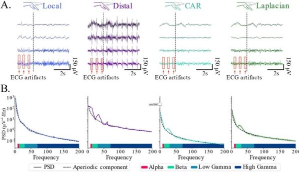
Carnegie Mellon Team Strips ECG Noise From Stentrode, Keeps Motor Signals

Stentrode, an endovascular brain-computer interface, often picks up electrocardiogram (ECG) artifacts. Researchers at Carnegie Mellon University and colleagues found that conventional re-referencing schemes reduce ECG artifacts but also diminish beta-band activity linked to movement. They applied band-limited independent component analysis (BL-ICA) as a spatial filter to remove ECG artifacts while preserving motor features. The study appeared in Advanced Science.
August 2026

Endovascular Electrodes Evoke Cortical Responses

The study was published in Journal of neural engineering on August 13, 2026, presenting the first strength-duration characterization of cortical evoked potentials elicited by endovascular stimulation adjacent to the cerebellum. The authors note that electrical stimulation and neural recording underpin neural prostheses for restoring function and treating neurological disorders, but that clinical adoption is limited by the invasiveness of implantation, while the Endovascular Neural Interface offers an alternative by accessing intracranial targets through the cerebral vasculature. A polymer-based stent-electrode array was deployed into the left transverse sinus of an ovine model, and biphasic current pulses targeting the cerebellum were delivered via the stent electrodes while a subdural electrocorticography grid recorded cortical responses. Endovascular stimulation consistently evoked time-locked cortical potentials with early and late components at approximately 40 ms and 100 ms post-stimulation, and impedance monitoring confirmed electrode functionality and stability throughout. Strength-duration analysis revealed rheobase and chronaxie values, providing a quantitative basis for parameter selection and comparison with established intracranial stimulation modalities.
© 2026 BCIwiki.com Digest Topics Tips Subscribe Revisions About